01Selected projects
Selfscope
At-Home Dermatoscope
Overview
01The product
A Dermatoscope for the Home
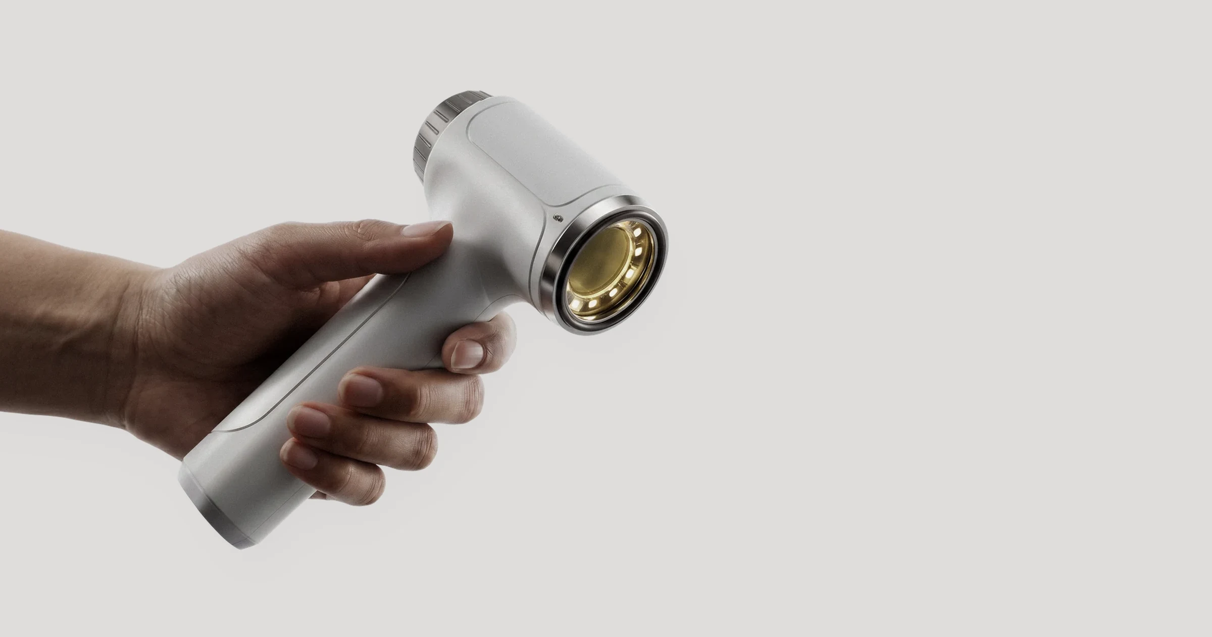
Selfscope is a dedicated at-home dermatoscope and companion interface that captures consistent images of skin lesions for review by a dermatologist.
It is designed for the whole household rather than the clinic: affordable and simple to use, without giving up the image quality and consistency that professional evaluation depends on. Selfscope does not diagnose. A dermatologist remains responsible for every assessment.
02The challenge
Skin Checks Are Still Rare
Australia has the highest incidence of skin cancer in the world (Wang et al., 2025). Caught early, most skin cancers are highly treatable, yet regular skin checks are not part of most people’s routine. A first check means living close enough to a clinic, paying for an appointment and making the time.
The options for an everyday person sit at two extremes. Phone apps and attachments are inexpensive but depend on the camera in each phone, so images of the same lesion are rarely consistent. Professional dermatoscopes are reliable, but are designed and priced for clinicians.
1Place
The silicone ring rests flat on intact skin and the illumination type is selected.
2Capture
A single press of the capture button records the image.
3Transfer
The image transfers to the user’s device, where it is placed on a body map for later professional review.
Design
03Form and ergonomics
Simple Enough to Keep at Home
The external form was designed so that it never suggests expert knowledge is needed to use it. Its smooth, uncluttered shape avoids the rows of buttons, dials and lights common on clinical equipment.
The head is angled at 10°, after testing showed that a straight form placed noticeably more tension on the wrist. The two controls sit on top of the handle rather than under the fingers like a drill trigger, where accidental presses were too easy.
Matte white ABS is paired with a stainless-steel front cap, communicating cleanliness, quality and precision.
04Illumination
Two Kinds of Light, One Button
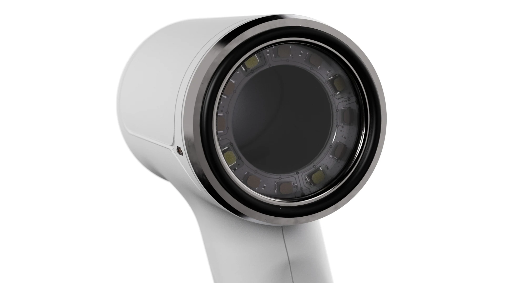
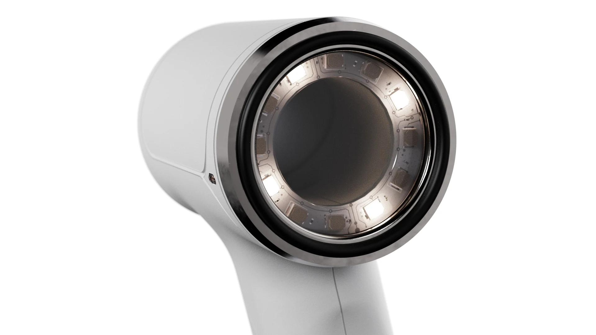
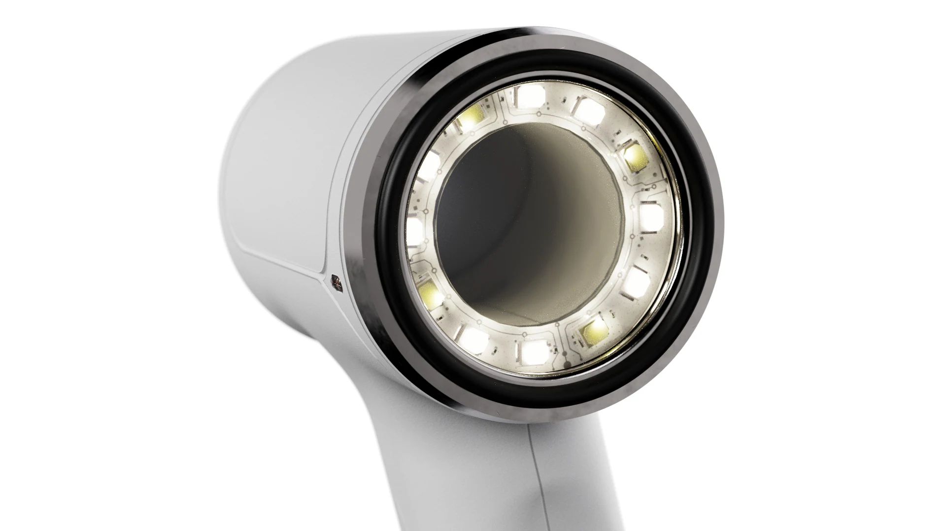
Light comes from a calibrated ring of 12 LEDs on a custom PCB, arranged around the optical axis to light the lesion evenly. Eight sit behind a polarising filter and four are uncovered, giving two modes alongside off.
Exposure and white balance are fixed, so images of the same lesion stay comparable over time.
Inside
05Optical path
What the User Sees Is What Is Captured
Light from the LED ring reflects off the skin and reaches a 50:50 beam splitter, which passes part of it straight through to the user’s eye and reflects part of it into the camera lens. One fixed path serves both.
The layout was worked out through digital ray simulation in 3DOptix before any physical testing. Simulating more than 20 configurations across six candidate lenses allowed the system to be simplified from three separate lenses to a single camera lens unit.
06Electronics and assembly
Four Boards, One System
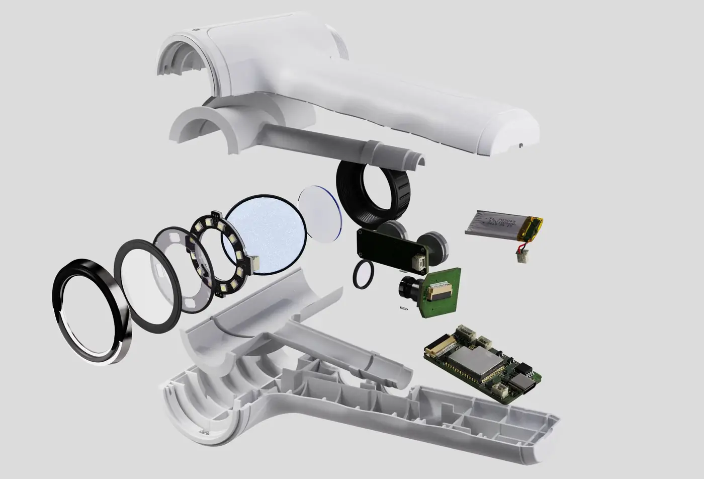
The electronics are divided across four custom boards: the LED ring, a dual-button board, an OV5640 5 MP camera board, and an MCU and power board built around an ESP32-S3 with USB-C charging and Bluetooth.
Scroll to take it apart
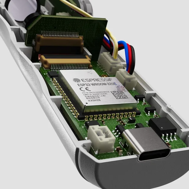
MCU and power PCBESP32-S3, battery charger and USB-C on 25.5 × 45.5 mm, with Bluetooth.
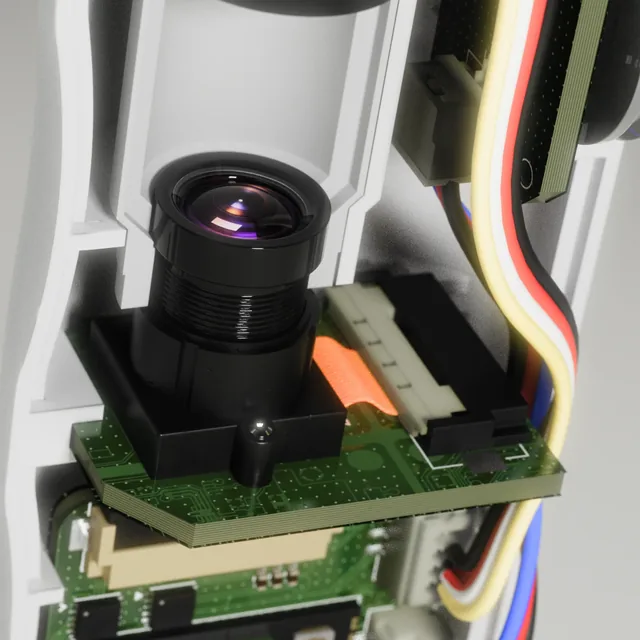
Camera systemOV5640 5 MP sensor behind a fixed M12, 12 mm lens.5 MPDedicated OV5640 camera sensor
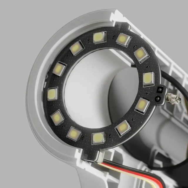
LED-ring PCB12 LEDs in two groups, one for each light mode.
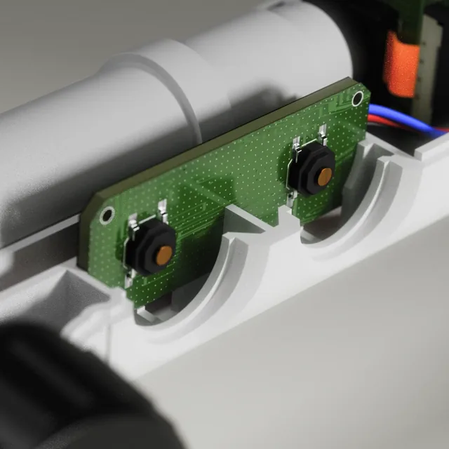
Button PCB and capsTwo tactile switches on a board of about 38 × 15.5 mm.
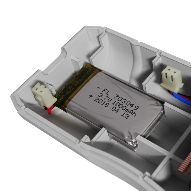
LiPo batteryProtected 3.7 V, 1000 mAh lithium cell.12 to 24Estimated check sessions per charge
System
07Companion interface
Images Live on the User’s Own Device
Images are viewed and organised in a companion interface rather than on the device, which removes the cost of a screen. Each capture is placed on a body map with a pin, and repeat captures of the same lesion are grouped so changes can be compared over time.
Store and Forward
The intended route to a dermatologist follows the store-and-forward model already used in teledermatology. Images stay on the user’s own device until they choose to send them for review.
Owned by the User
The user owns the images, can delete them at any time, and sees who has viewed them, reflecting the protections the Privacy Act 1988 (Cth) gives health information.
08Ownership package
A Complete Package, Not a Device Alone
A fitted EVA case with a foam insert protects Selfscope during storage and transport, and a mesh pocket in the lid holds the printed documentation.
The documentation consists of a 32-page A5 Instructions for Use, an A6 Quick Start Guide and a device identification label. The Quick Start Guide reduces normal use to six steps, so the full manual is not needed day to day.
Printed documentation
Click or drag a page to turn it
Instructions for UseA5, 32 pages
Quick Start GuideA6, double-sided
Process
09Development
From Printed Forms to Working Electronics
The form was refined through printed iterations before the optics and electronics were brought together in a working build.
10Prototype
The Working Prototype
The prototype proves that the components, PCBs and optical layout fit and work together. It was shown with an exploded view model, the case and the manuals.
11Outcome
Selfscope gives a household a simple, affordable way to capture consistent images of skin lesions for a dermatologist to review. The project ran from printed form studies and optical simulation through to a working prototype with custom electronics and a companion interface.
When the user isn’t an expert, the expertise has to be built into the object.